Retinal laser (argon)


Retinal foramen
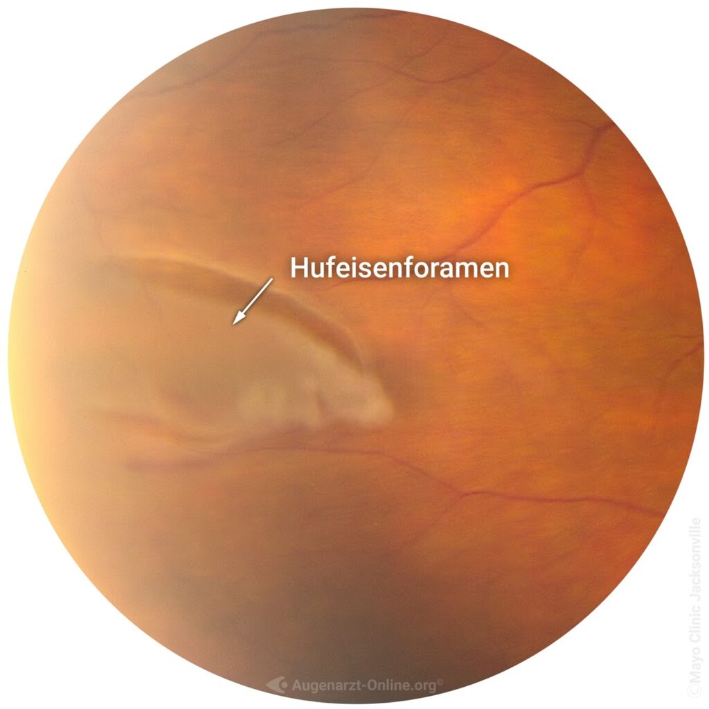
If you see flashes, black shadows or a so-called soot rain, then an acute examination of the retina should be carried out. These symptoms may indicate a change in the vitreous body. The vitreous body is located inside the eye and detaches from the retina in the course of life. In rare cases, the vitreous body can cause tears in the retina during this process (retinal foramen) (Fig. 1). If such a retinal foramen develops, it must be treated to minimize the risk of retinal detachment. Retinal foramen are treated with the argon laser in which the tear is surrounded circularly with laser foci and the retina is thus "welded" to the underlying choroid/sclera.
Which people are particularly at risk of a retinal foramen?
Patients with the following characteristics have a higher risk of developing a retinal tear:
- Degenerative zones in the peripheral retina
- Thin retinal areas (mostly in the periphery)
- Premature vitreous detachment (usually as a result of trauma to the eyeball)
- Patients with higher myopia
Diabetes laser treatment
What is diabetic retinopathy?
The eye and the retina are one of the organs with the best blood supply and react very sensitively to reduced blood flow. Diabetes can cause lasting damage to the small vessels of the retina and therefore also to the sensory cells.
What is vascular damage?
The vessel walls of these small vessels can become brittle and leaky as a result of elevated blood sugar levels, which have usually been present for many years, as well as increased blood pressure and elevated blood lipids. If the vessels are leaking, blood and fluid or even fatty bodies leak into the retina, which the ophthalmologist can discover during fundoscopy. (Image 1b)
What are the consequences of vascular damage for vision?
As the vascular changes are painless, they are only noticed by those affected when their vision is severely impaired.
Therefore, patients usually do not notice the changes in the early stages. Only early detection by an ophthalmologist can prevent visual deterioration for as long as possible.
What types of changes are there?
1. diabetic retinopathy
This refers to the disease of the peripheral retina. With advanced disease, chronic reduced blood flow can lead to functional deficits in large areas of the retina. If this is not treated, it can lead to serious complications such as the formation of new blood vessels, bleeding (Fig. 1b) into the interior of the eye or even retinal detachment.
2. diabetic maculopathy
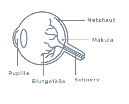
The macula is located in the center of the retina (image 2a). It is responsible for visual acuity and enables us to read, drive, see objects and colors in detail and recognize faces.
In diabetic maculopathy, initially only small hemorrhages appear, which the patient usually barely notices. Subsequently, fluid leaks into the retina through "leaky vessels" and swelling occurs in the area of the central macula. This swelling (Fig. 2b) is associated with a deterioration in vision (Fig. 2d) and causes damage to the photoreceptors, meaning that permanent damage can occur if treatment is not carried out.
For a precise assessment of progressive disease or the appearance of fluid, we recommend carrying out optical coherence tomography (OCT - Figures 2a and 2b).


What treatment options are there for diabetic maculopathy?
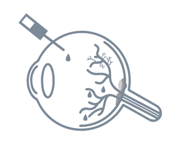
If there is fluid in the central macula, so-called anti-VEGF or corticosteroids are administered into the eye using IVOM. This therapy leads to a reduction in fluid leakage from the damaged vessels and thus to a stabilization and usually also to an improvement in the findings.
We will be happy to advise you on the latest literature and therapeutic guidelines.
Which therapiesöWhat are the options for diabetic retinopathy?
Targeted laser therapy is used if the disease is severe. This treats areas of the retina that are already damaged and non-functional, thus protecting the visual center in the long term.
At the Angermann Eye Center, we will advise you on all the options and look for the best solution for you and, if necessary, can perform the laser on our premises
Can diabetes cause blindness?
There is a risk of blindness if you are not careful, but this can be significantly reduced by regular check-ups with an ophthalmologist. Cooperation with an internist is also of crucial importance. Lowering blood pressure, blood lipids and long-term glucose (HbA1c) to at least below 7.5% significantly reduces the risk of complications!
Prevention is the best therapy. Because only if the changes are detected in good time can the right treatment be initiated.
Our recommendations:
- Six-monthly check-ups at the ophthalmologist - depending on the findings, check-ups or treatments may also be necessary every 3 months or more frequently.
- Once a year examination with dilation of the pupil.
- Immediate re-presentation in the event of a sudden deterioration in vision.
- OCT examination for abnormal findings
Diabetes is a metabolic disorder that leads to increased blood sugar levels. In diabetic retinopathy, the vessels in the retina become diseased as a result of the metabolic disorder. In laser therapy - also known as argon laser coagulation - targeted laser beams are directed at the damaged retina, destroying the pathological proliferation of blood vessels and suppressing the formation of further vascular changes, among other things. Laser treatment is not sufficient in cases of advanced disease with extensive vascular proliferation and severe bleeding inside the eye.
Argon laser coagulation (ALK) is a procedure used to treat various forms of diabetic eye disease. It involves destroying abnormally proliferating blood vessels on the retina using a focused beam of light. The laser is also used to create tiny scars (0.01-0.02mm) around diseased tissue to prevent it from spreading. This leads to an improvement in the oxygen supply to the macula (the point of sharpest vision in the human eye) and, as a result, to a stabilization or improvement in visual acuity.
What is diabetic retinopathy?
Diabetic retinopathy is a disease of the retina caused by diabetes mellitus. Due to metabolic disorders, the vessels of the retina become diseased and microaneurysms, hemorrhages, fatty deposits and microinfarcts of the retina can occur. Diabetic retinopathy is the most common cause of blindness worldwide in people between the ages of 20 and 60.
Prevention and alternative treatment methods
As an alternative to ALK treatment, diabetic retinopathy can also be treated with injection therapy or surgery. Both in the prevention and treatment of diabetic retinopathy, it is extremely important to always keep an eye on blood glucose levels and to adjust them correctly. As a general rule, no form of treatment has any prospect of long-term success if the diabetes causing the eye disease is not treated.

